SIALENDOSCOPY
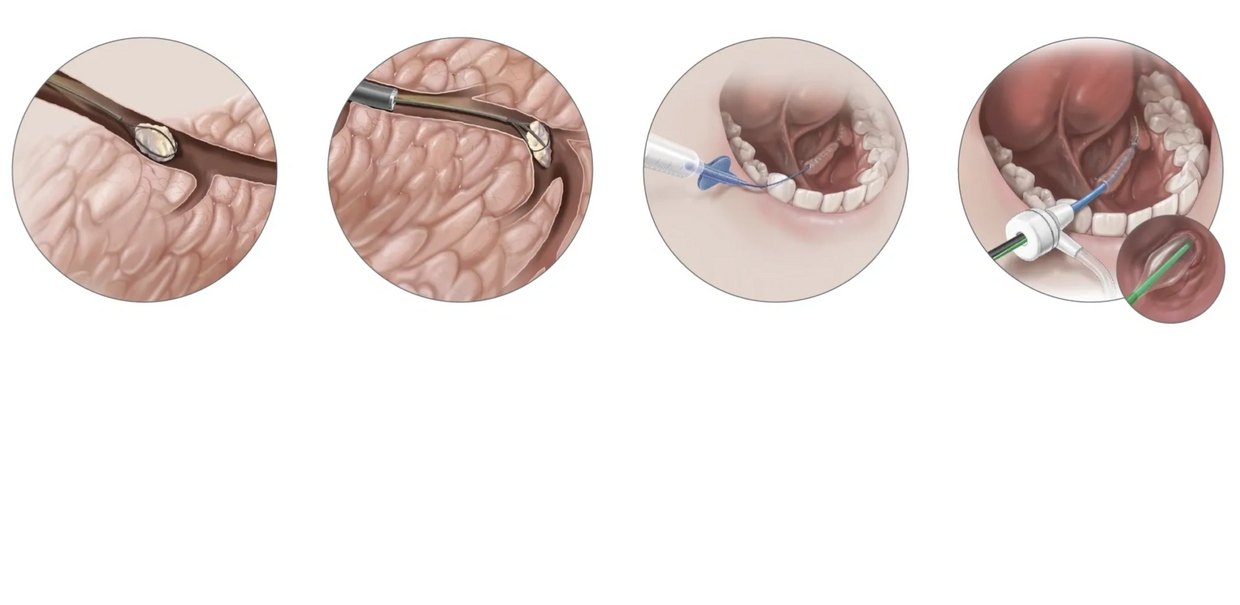
Sialendoscopy has emerged as the method of choice for treatment of sialoliths (salivary gland stones).
In obstructive salivary pathologies, sialendoscopy is currently being used largely for therapeutic purposes and the first diagnostic tool of investigation.
Availability of new miniaturized instruments for therapeutic purposes and enhanced optical resolution has increase the efficacy and precision of sialendoscopy in the management of salivary gland pathologies.
Sialendoscopy has emerged as a preferred diagnostic as well as therapeutic tool for management of salivary gland pathologies and has helped significantly reduce the morbidity, loss of work hours and hospital stay.
Advantages -
- Sialendoscope is a good diagnostic tool for ductal pathology and unlike other radiological procedures findings of sialendoscopy correlate fairly with the symptomatology.
- Sialendoscopy can be used both for diagnostic as well as therapeutic purposes and very often it can be done in a single sitting. Though it is also an invasive procedure, morbidity associated with Sialendoscopy is mostly minor and that too most of the time is temporary.
Indications
Currently, Sialendoscopy is being most commonly used for removing submandibular and parotid duct calculi which is the most common obstructive pathology affecting the salivary glands.
Earlier, salivary duct stones were being managed surgically, either by removing the entire salivary gland or by marsupializing the duct and removal of the stone. But Stenson’s duct stones were a real challenge. Though parotidectomy was advised for treatment of parotid stones, it was rarely performed, due to inherent risk of facial nerve injury. Thus, most of these patients used to remain untreated and would suffer from recurrent parotitis. Sialendoscope has brought a paradigm shift in management of parotid duct stones. Stones can be removed through the sialendoscope itself. Alternatively, Large stones can be removed with combined approach (sialendoscope guided external approach).
Juvenile recurrent parotitis (JRP) is recurrent inflammatory condition of the parotid glands affecting the paediatric population, with unknown aetiology. Sialendoscopy in these patients helps in understanding the pathology, ruling out any localized cause and it has a therapeutic role also. Steroids which are being used systemically for this pathology can now be delivered directly to the desired site, through the scope. This increases the efficacy and reduces the systemic side effects of the steroid treatment, reflecting in overall improved control and/or cure rate.
Ductal strictures which may be secondary to ductal calculus can be very effectively treated with the help of sialendoscopes. Sialendoscopy helps in direct and precise evaluation of the nature, site and length of the ductal strictures and consequently the optimal intervention.
This website uses cookies.
We use cookies to analyze website traffic and optimize your website experience. By accepting our use of cookies, your data will be aggregated with all other user data.
